 Sensory-Tactile Functional Mapping and Use-Associated Structural Variation of the Human Female Genital Representation Field | Journal of Neuroscience
https://www.jneurosci.org/content/early/2021/12/09/JNEUROSCI.1081-21.2021
Sensory-Tactile Functional Mapping and Use-Associated Structural Variation of the Human Female Genital Representation Field
Abstract
The precise location of the human female genital representation field in the primary somatosensory cortex (S1) is controversial and its capacity for use-associated structural variation as a function of sexual behavior remains unknown. We used an fMRI-compatible sensory-tactile stimulation paradigm to functionally map the location of the female genital representation field in 20 adult women. Neural response to tactile stimulation of the clitoral region (versus right hand) identified individually-diverse focal bilateral activations in dorsolateral areas of S1 (BA1-BA3) in alignment with anatomical location. We next used cortical surface analyses to assess structural thickness across the 10 individually most activated vertices per hemisphere for each woman. We show that frequency of sexual intercourse within 12 months is correlated with structural thickness of the individually-mapped left genital field. Our results provide a precise functional localization of the female genital field and provide support for use-associated structural variation of the human genital cortex.
SIGNIFCANCE
We provide a precise location of the human female genital field in the somatosensory cortex and, for the first time, provide evidence in support of structural variation of the human genital field in association with frequency of genital contact. Our study represents a significant methodological advance by individually mapping genital fields for structural analyses. On a secondary level, our results suggest that any study investigating changes in the human genital field must map the field individually to achieve sufficient precision. Our results pave the way for future research into the plasticity of the human genital cortex as a function of normal or adverse experience as well as changes in pathological conditions, i.e. sexual dysfunction, sexual deviation or sexual risk-taking behavior.
Funded by a NeuroCure Cluster of Excellence (Deutsche Forschungsgemeinschaft EXC 2049) intramural innovation grant to CH and MB, the Max Planck School of Cognition to CH and JDH, and a scholarship from the Einstein Center for Neuroscience Berlin to AJJK. We thank Kristina Sandt and Frieda Born for assistance.
世界で初めて「クリトリスへの刺激」に対応する脳領域がマッピングされる

性的な快感は、神経から受け取った信号を脳で処理する報酬系によって得られるといわれていますが、具体的にどの脳領域で処理されているのかは明らかになっていません。ドイツ・ベルリンにあるシャリテ大学病院の研究チームによる研究で、女性器への接触に関連する脳領域が機能的磁気共鳴画像法(fMRI)により特定され、セックスの経験回数が多い人ほどその脳領域が発達していることが判明しました。
Sensory-Tactile Functional Mapping and Use-Associated Structural Variation of the Human Female Genital Representation Field | Journal of Neuroscience
https://www.jneurosci.org/content/early/2021/12/09/JNEUROSCI.1081-21.2021
For The First Time, Scientists Map Brain Regions Responding to The Clitoris
https://www.sciencealert.com/science-has-located-the-brain-region-that-responds-to-the-clitoris
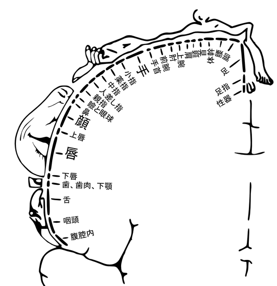
触覚や温度感覚、痛覚など、体全体の皮膚感覚情報を受信して処理するのが大脳にある体性感覚皮質です。体の各部位の体性感覚は体性感覚皮質の異なる領域に対応しており、研究によって少しずつマッピングが行われています。

女性器の体性感覚がどの領域で行われているのかについてはこれまで議論が行われており、足裏と同じ部位だとする説や、腰に近い部位だとする説が唱えられました。ただし、女性器への刺激は自分あるいはパートナーの指によるものだったため、体の他の部位が同時に触られたり刺激方法が不正確だったりするため、結果の信頼性は低いのが問題でした。
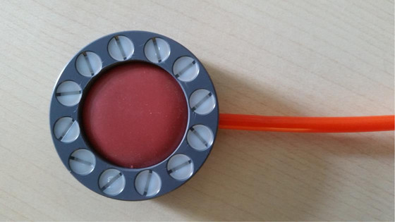
そこで、今回の実験では以下の装置が使われました。この装置を下着の上からクリトリスの高さに当てると、赤く丸い幕部分が空気の噴射によって小さく振動するという仕組みで、クリトリスに狙いを定めて刺激を与えることができます。シャリテ大学病院のベルリン先端神経画像センターの研究員であるジョン=ディラン・ヘインズ氏は、この装置が被験者にとって「できるだけ快適」であるように設計されたものだと述べています。

実験では、18から45歳までの健康な女性20人が、fMRIによって脳部位の活性化を監視されながら、10秒間の休憩をはさみつつ、10秒間×8回の刺激を受けました。対照として、同じ装置を右手の甲にも使用したとのこと。
その結果、女性器と対応した体性感覚皮質領域は、男性と同じように腰の近くにあることが判明しましたが、正確な位置は被験者によって異なりました。また、性交の回数が多いほど、女性器と対応する領域は大きくなることもわかりました。

脳の特定部位を使えば使うほどその部位が大きくなるという「脳の可塑性」はこれまでにも言及されています。過去の研究では、ラットとマウスへの生殖器の刺激で脳の特定領域が大きくなっていることもわかっています。ただし、2013年の研究では、外傷性の性的暴力に苦しんでいる人の脳内では、性器に対応した脳領域が小さくなる可能性が示唆されています。
シャリテ大学の医療心理学教授であるクリスティン・ハイム氏は、「女性器と対応した脳領域を特定すること、そして女性の性的経験に関連してどのように変化するかは未解明です。今回の実験結果は将来的に、性的暴力の影響を受けたり性的機能不全に陥ったりした人たちの治療に応用できるかもしれません」とコメントしました。
0 件のコメント:
コメントを投稿